
International
ADVANCED AND APPLIED SCIENCES
EISSN: 2313-3724, Print ISSN: 2313-626X
Frequency: 12
![]()
Volume 10, Issue 8 (August 2023), Pages: 51-63

----------------------------------------------
Original Research Paper
Advances in neonatal brain imaging: A comparative analysis of MRI, CT scans, and ultrasound
Author(s):
Arwa O. Baeshen 1, Naif H. Almutairi 1, Othman I. Alomair 1, Dhafer M. Alahmari 2, Magbool Alelyani 3, Sami A. Alghamdi 1, *
Affiliation(s):
1Department of Radiological Sciences, College of Applied Medical Sciences, King Saud University, Riyadh, Saudi Arabia
2Department of Medical Imaging, King Saud Medical City, Riyadh, Saudi Arabia
3Department of Radiological Sciences, College of Applied Medical Sciences, King Khalid University, Abha, Saudi Arabia

* Corresponding Author.
 Corresponding author's ORCID profile: https://orcid.org/0000-0002-6404-8762
Digital Object Identifier:
https://doi.org/10.21833/ijaas.2023.08.006
Abstract:
This scholarly investigation undertakes a comprehensive comparison of the diagnostic efficacy, precision, and sensitivity associated with neonatal brain Magnetic Resonance Imaging (MRI) in contrast to its counterparts, Computed Tomography (CT) scans and ultrasound. As the medical community has progressively become attuned to the long-term health implications of radiation exposure from CT scans, the imperative of a strategy mitigating this risk has gained prominence. In this context, ultrasound emerges as an alternative modality devoid of ionizing radiation. Employing a methodical approach rooted in systematic literature review, this study synthesizes five pertinent research works to unravel its research objectives. Empirical evidence substantiates that neonatal brain MRI surpasses both neonatal brain CT and ultrasound in diagnostic effectiveness. The underpinning rationale for this phenomenon lies in the heightened accuracy inherent to neonatal brain MRI procedures. To unravel the intricacies associated with disparities between neonatal and adult brain MRI procedures, the study meticulously investigates structural, shape, and size distinctions. This endeavor underscores the necessity for bespoke MRI apparatuses designed to account for these nuances. In pursuit of this objective, the integration of technologically advanced components such as compact scanners and refinements in magnetic and coil technologies has engendered tangible improvements. This innovation confluence bears testimony to the augmentation of patient safety, conferring a cascade effect wherein the precision of acquired MRI data underwrites accurate diagnoses and consequent therapeutic interventions. Conclusively, the study underscores the pivotal role played by recent MRI technological advancements in amplifying its efficacy within the niche domain of neonatal brain imaging. Prospective innovations within the MRI ambit stand poised to recalibrate performance benchmarks, thereby amplifying its diagnostic potency and broadening its scope of application.
© 2023 The Authors. Published by IASE.
This is an
Keywords: Neonatal brain MRI, CT scan, Ultrasound, Radiation exposure, Technological advancements
Article History: Received 26 February 2023, Received in revised form 20 June 2023, Accepted 25 June 2023
Acknowledgment
No Acknowledgment.
Compliance with ethical standards
Conflict of interest: The author(s) declared no potential conflicts of interest with respect to the research, authorship, and/or publication of this article.
Citation:
Baeshen AO, Almutairi NH, Alomair OI, Alahmari DM, Alelyani M, and Alghamdi SA (2023). Advances in neonatal brain imaging: A comparative analysis of MRI, CT scans, and ultrasound. International Journal of Advanced and Applied Sciences, 10(8): 51-63
Figures
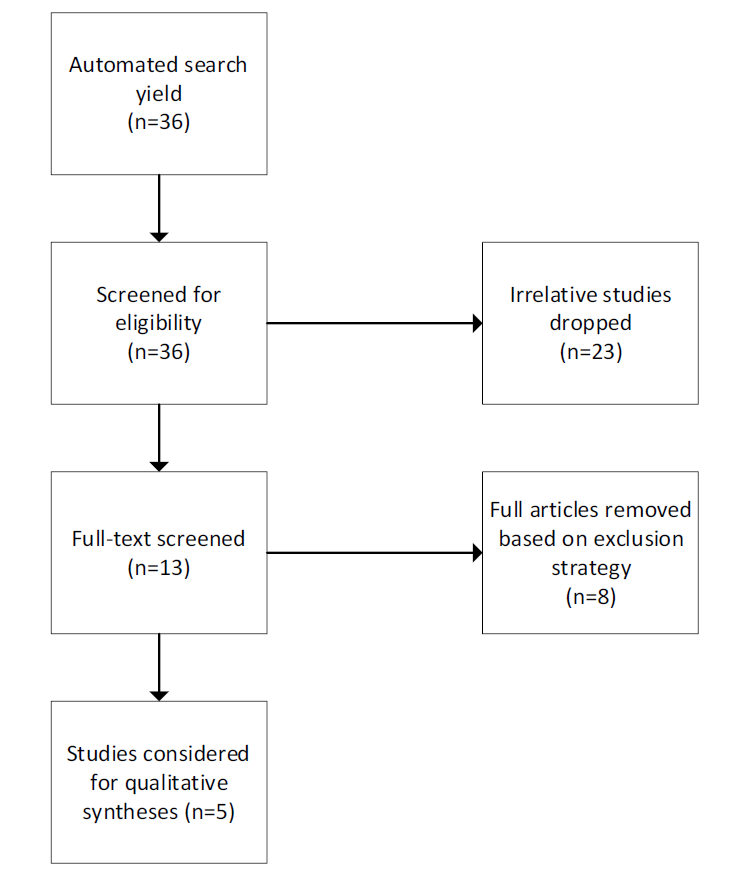
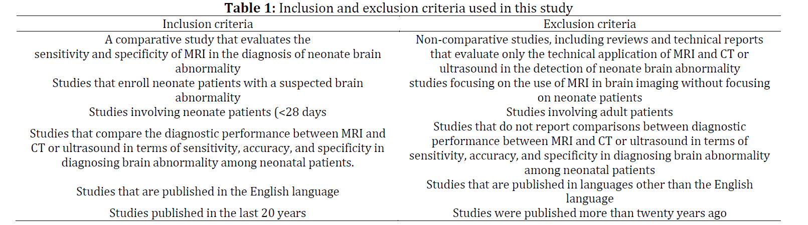
Tables
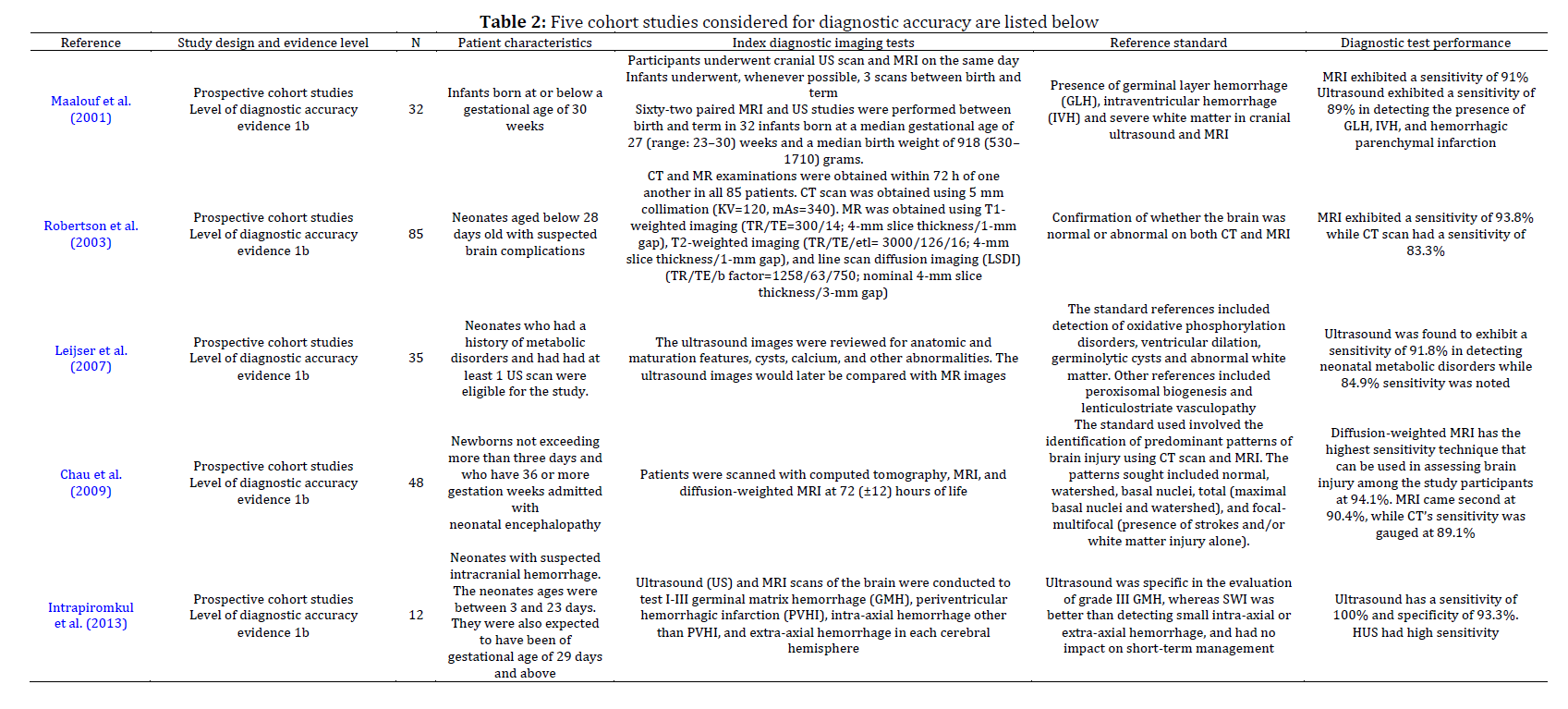
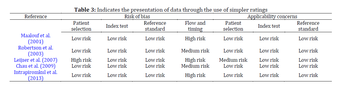
----------------------------------------------
References (44)
- Ahmad SF, Kim YC, Choi IC, and Kim HD (2020). Recent progress in birdcage RF coil technology for MRI system. Diagnostics, 10(12): 1017. https://doi.org/10.3390/diagnostics10121017 [Google Scholar] PMid:33261167 PMCid:PMC7759766
- Alkalay AL, Flores-Sarnat L, Sarnat HB, Moser FG, and Simmons CF (2005). Brain imaging findings in neonatal hypoglycemia: Case report and review of 23 cases. Clinical Pediatrics, 44(9): 783-790. https://doi.org/10.1177/000992280504400906 [Google Scholar] PMid:16327965
- Audrey S and Procter S (2015). Employers’ views of promoting walking to work: A qualitative study. International Journal of Behavioral Nutrition and Physical Activity, 12: 12. https://doi.org/10.1186/s12966-015-0174-8 [Google Scholar] PMid:25888840 PMCid:PMC4344752
- Barkovich AJ, Miller SP, Bartha A, Newton N, Hamrick SEG, Mukherjee P, and Vigneron DB (2006). MR imaging, MR spectroscopy, and diffusion tensor imaging of sequential studies in neonates with encephalopathy. American Journal of Neuroradiology, 27(3): 533-547. [Google Scholar]
- Barnette AR, Horbar JD, Soll RF, Pfister RH, Nelson KB, Kenny MJ, and Inder TE (2014). Neuroimaging in the evaluation of neonatal encephalopathy. Pediatrics, 133(6): e1508-e1517. https://doi.org/10.1542/peds.2013-4247 [Google Scholar] PMid:24864165
- Bekiesińska-Figatowska M, Rutkowska M, Stankiewicz J, Krupa K, Iwanowska B, Romaniuk-Doroszewska A, and Helwich E (2019). Neonatal brain and body imaging in the MR-compatible incubator. Advances in Clinical and Experimental Medicine, 28(7): 945-954. https://doi.org/10.17219/acem/94155 [Google Scholar] PMid:31111693
- Blamire AM (2008). The technology of MRI-The next 10 years? The British Journal of Radiology, 81(968): 601-617. https://doi.org/10.1259/bjr/96872829 [Google Scholar] PMid:18628329
- Buehrer M, Pruessmann KP, Boesiger P, and Kozerke S (2007). Array compression for MRI with large coil arrays. Magnetic Resonance in Medicine: An Official Journal of the International Society for Magnetic Resonance in Medicine, 57(6): 1131-1139. https://doi.org/10.1002/mrm.21237 [Google Scholar] PMid:17534913
- Chartier AL, Bouvier MJ, McPherson DR, Stepenosky JE, Taysom DA, and Marks RM (2019). The safety of maternal and fetal MRI at 3 T. American Journal of Roentgenology, 213(5): 1170-1173. https://doi.org/10.2214/AJR.19.21400 [Google Scholar] PMid:31310182
- Chau V, Poskitt KJ, Sargent MA, Lupton BA, Hill A, Roland E, and Miller SP (2009). Comparison of computer tomography and magnetic resonance imaging scans on the third day of life in term newborns with neonatal encephalopathy. Pediatrics, 123(1): 319-326. https://doi.org/10.1542/peds.2008-0283 [Google Scholar] PMid:19117898
- Devi CN, Chandrasekharan A, Sundararaman VK, and Alex ZC (2015). Neonatal brain MRI segmentation: A review. Computers in Biology and Medicine, 64: 163-178. https://doi.org/10.1016/j.compbiomed.2015.06.016 [Google Scholar] PMid:26189155
- Dubois J, Alison M, Counsell SJ, Hertz‐Pannier L, Hüppi PS, and Benders MJ (2021). MRI of the neonatal brain: A review of methodological challenges and neuroscientific advances. Journal of Magnetic Resonance Imaging, 53(5): 1318-1343. https://doi.org/10.1002/jmri.27192 [Google Scholar] PMid:32420684 PMCid:PMC8247362
- Dudink J, Jeanne Steggerda S, and Horsch S (2020). State-of-the-art neonatal cerebral ultrasound: Technique and reporting. Pediatric Research, 87(Suppl 1): 3-12. https://doi.org/10.1038/s41390-020-0776-y [Google Scholar] PMid:32218539 PMCid:PMC7098885
- Duftner C, Dejaco C, Sepriano A, Falzon L, Schmidt WA, and Ramiro S (2018). Imaging in diagnosis, outcome prediction and monitoring of large vessel vasculitis: A systematic literature review and meta-analysis informing the EULAR recommendations. RMD Open, 4: e000612. https://doi.org/10.1136/rmdopen-2017-000612 [Google Scholar] PMid:29531788 PMCid:PMC5845406
- Fumagalli M, Cinnante CM, Calloni SF, Sorrentino G, Gorla I, Plevani L, and Triulzi F (2018). Clinical safety of 3-T brain magnetic resonance imaging in newborns. Pediatric Radiology, 48: 992-998. https://doi.org/10.1007/s00247-018-4105-0 [Google Scholar] PMid:29594440
- Gannon CM, Kornhauser MS, Gross GW, Wiswell TE, Baumgart S, Streletz LJ, and Spitzer AR (2001). When combined, early bedside head ultrasound and electroencephalography predict abnormal computerized tomography or magnetic resonance brain images obtained after extracorporeal membrane oxygenation treatment. Journal of Perinatology, 21(7): 451-455. https://doi.org/10.1038/sj.jp.7210593 [Google Scholar] PMid:11894513
- Genedi EAS, Osman NM, and El-deeb MT (2016). Magnetic resonance imaging versus transcranial ultrasound in early identification of cerebral injuries in neonatal encephalopathy. The Egyptian Journal of Radiology and Nuclear Medicine, 47(1): 297-304. https://doi.org/10.1016/j.ejrnm.2016.01.001 [Google Scholar]
- Groenendaal F and de Vries LS (2017). Fifty years of brain imaging in neonatal encephalopathy following perinatal asphyxia. Pediatric Research, 81(1): 150-155. https://doi.org/10.1038/pr.2016.195 [Google Scholar] PMid:27673422
- Harvey ME and Redshaw ME (2016). Qualitative study of the clinician–parent interface in discussing prognosis following MRI and US imaging of preterm infants in the UK. BMJ Open, 6(9): e011472. https://doi.org/10.1136/bmjopen-2016-011472 [Google Scholar] PMid:27678531 PMCid:PMC5051465
- Hughes EJ, Winchman T, Padormo F, Teixeira R, Wurie J, Sharma M, and Hajnal JV (2017). A dedicated neonatal brain imaging system. Magnetic Resonance in Medicine, 78(2): 794-804. https://doi.org/10.1002/mrm.26462 [Google Scholar] PMid:27643791 PMCid:PMC5516134
- Ibrahim J, Mir I, and Chalak L (2018). Brain imaging in preterm infants< 32 weeks gestation: A clinical review and algorithm for the use of cranial ultrasound and qualitative brain MRI. Pediatric Research, 84(6): 799-806. https://doi.org/10.1038/s41390-018-0194-6 [Google Scholar] PMid:30315272
- Intrapiromkul J, Northington F, Huisman TA, Izbudak I, Meoded A, and Tekes A (2013). Accuracy of head ultrasound for the detection of intracranial hemorrhage in preterm neonates: Comparison with brain MRI and susceptibility-weighted imaging. Journal of Neuroradiology, 40(2): 81-88. https://doi.org/10.1016/j.neurad.2012.03.006 [Google Scholar] PMid:22633043 PMCid:PMC4428334
- Khan IA (2021). Do second generation sequencing techniques identify documented genetic markers for neonatal diabetes mellitus? Heliyon, 7(9): e07903. https://doi.org/10.1016/j.heliyon.2021.e07903 [Google Scholar] PMid:34584998 PMCid:PMC8455689
- Lane A, Chuk LMR, Colditz PB, and Coulthard A (2013). The MRI‐compatible neonatal incubator in practice. Journal of Paediatrics and Child Health, 49(9): E377-E380. https://doi.org/10.1111/jpc.12222 [Google Scholar] PMid:23678957
- Leijser LM, De Vries LS, Rutherford MA, Manzur AY, Groenendaal F, De Koning TJ, and Cowan FM (2007). Cranial ultrasound in metabolic disorders presenting in the neonatal period: Characteristic features and comparison with MR imaging. American Journal of Neuroradiology, 28(7): 1223-1231. https://doi.org/10.3174/ajnr.A0553 [Google Scholar] PMid:17698520 PMCid:PMC7977655
- Li G, Wang L, Yap PT, Wang F, Wu Z, Meng Y, and Shen D (2019). Computational neuroanatomy of baby brains: A review. NeuroImage, 185: 906-925. https://doi.org/10.1016/j.neuroimage.2018.03.042 [Google Scholar] PMid:29574033 PMCid:PMC6150852
- Likeman M (2014). MRI brain imaging in neonates. Paediatrics and Child Health, 24(9): 407-412. https://doi.org/10.1016/j.paed.2014.02.005 [Google Scholar]
- Lindberg DM, Stence NV, Grubenhoff JA, Lewis T, Mirsky DM, Miller AL, and Runyan DK (2019). Feasibility and accuracy of fast MRI versus CT for traumatic brain injury in young children. Pediatrics, 144(4): e20190419. https://doi.org/10.1542/peds.2019-0419 [Google Scholar] PMid:31533974
- Maalouf EF, Duggan PJ, Counsell SJ, Rutherford MA, Cowan F, Azzopardi D, and Edwards AD (2001). Comparison of findings on cranial ultrasound and magnetic resonance imaging in preterm infants. Pediatrics, 107(4): 719-727. https://doi.org/10.1542/peds.107.4.719 [Google Scholar] PMid:11335750
- Malamateniou C, Malik SJ, Counsell SJ, Allsop JM, McGuinness AK, Hayat T, and Rutherford MA (2013). Motion-compensation techniques in neonatal and fetal MR imaging. American Journal of Neuroradiology, 34(6): 1124-1136. https://doi.org/10.3174/ajnr.A3128 [Google Scholar] PMid:22576885 PMCid:PMC7964586
- Nopoulos P, Berg S, Castellenos FX, Delgado A, Andreasen NC, and Rapoport JL (2000). Developmental brain anomalies in children with attention-deficit hyperactivity disorder. Journal of Child Neurology, 15(2): 102-108. https://doi.org/10.1177/088307380001500208 [Google Scholar] PMid:10695895
- Picone S, Ariganello P, Mondì V, Di Palma F, Martini L, Marziali S, and Paolillo P (2019). A solution based on melatonin, tryptophan, and vitamin B6 (Melamil Tripto©) for sedation in newborns during brain MRI. Italian Journal of Pediatrics, 45(1): 122. https://doi.org/10.1186/s13052-019-0714-y [Google Scholar] PMid:31547831 PMCid:PMC6757392
- Plaisier A, Raets MM, van der Starre C, Feijen-Roon M, Govaert P, Lequin MH, and Dudink J (2012). Safety of routine early MRI in preterm infants. Pediatric Radiology, 42: 1205-1211. https://doi.org/10.1007/s00247-012-2426-y [Google Scholar] PMid:22875205 PMCid:PMC3460174
- Prager A and Roychowdhury S (2007). Magnetic resonance imaging of the neonatal brain. The Indian Journal of Pediatrics, 74: 173-184. https://doi.org/10.1007/s12098-007-0012-3 [Google Scholar] PMid:17337831
- Robertson RL, Robson CD, Zurakowski D, Antiles S, Strauss K, and Mulkern RV (2003). CT versus MR in neonatal brain imaging at term. Pediatric Radiology, 33: 442-449. https://doi.org/10.1007/s00247-003-0933-6 [Google Scholar] PMid:12743660
- Saunders DE, Thompson C, Gunny R, Jones R, Cox T, and Chong WK (2007). Magnetic resonance imaging protocols for paediatric neuroradiology. Pediatric Radiology, 37: 789-797. https://doi.org/10.1007/s00247-007-0462-9 [Google Scholar] PMid:17487479 PMCid:PMC1950216
- Shi F, Fan Y, Tang S, Gilmore JH, Lin W, and Shen D (2010). Neonatal brain image segmentation in longitudinal MRI studies. Neuroimage, 49(1): 391-400. https://doi.org/10.1016/j.neuroimage.2009.07.066 [Google Scholar] PMid:19660558 PMCid:PMC2764995
- Smiljkovic M, Renaud C, Tapiero B, Lamarre V, and Kakkar F (2019). Head ultrasound, CT or MRI? The choice of neuroimaging in the assessment of infants with congenital cytomegalovirus infection. BMC Pediatrics, 19(1): 180. https://doi.org/10.1186/s12887-019-1562-z [Google Scholar] PMid:31167649 PMCid:PMC6549373
- Sorokan ST, Jefferies AL, and Miller SP (2018). Imaging the term neonatal brain. Paediatrics and Child Health, 23(5): 322-328. https://doi.org/10.1093/pch/pxx161 [Google Scholar] PMid:30657135 PMCid:PMC6054233
- Tkach JA, Hillman NH, Jobe AH, Loew W, Pratt RG, Daniels BR, and Dumoulin CL (2012). An MRI system for imaging neonates in the NICU: Initial feasibility study. Pediatric Radiology, 42: 1347-1356. https://doi.org/10.1007/s00247-012-2444-9 [Google Scholar] PMid:22735927
- Tkach JA, Merhar SL, Kline-Fath BM, Pratt RG, Loew WM, Daniels BR, and Dumoulin CL (2014). MRI in the neonatal ICU: Initial experience using a small-footprint 1.5-T system. American Journal of Roentgenology, 202(1): W95-W105. https://doi.org/10.2214/AJR.13.10613 [Google Scholar] PMid:24370170
- Weisenfeld NI and Warfield SK (2009). Automatic segmentation of newborn brain MRI. Neuroimage, 47(2): 564-572. https://doi.org/10.1016/j.neuroimage.2009.04.068 [Google Scholar] PMid:19409502 PMCid:PMC2945911
- Xiao Y and Watson M (2019). Guidance on conducting a systematic literature review. Journal of Planning Education and Research, 39(1): 93-112. https://doi.org/10.1177/0739456X17723971 [Google Scholar]
- Yu JC, Khodadadi H, Malik A, Davidson B, and Salles EDSL, Bhatia J, and Baban B (2018). Innate immunity of neonates and infants. Frontiers in Immunology, 9: 1759. https://doi.org/10.3389/fimmu.2018.01759 [Google Scholar] PMid:30105028 PMCid:PMC6077196